Deep in vivo imaging with super high-sensitive GaAsP NDD
The GaAsP NDD is equipped with GaAsP PMT that has a higher signal-to-noise ratio and superior sensitivity than a Multi-Alkali PMT, and allows clear imaging of deeper areas of living specimens. Its ability to acquire images of high S/N ratio, enables faster imaging and higher quality Z-stack imaging. Its high sensitivity allows the acquisition of fluorescent signals with less laser power, resulting in less photo damage to living specimens.
The Nikon A1 MP+ and A1R MP+ can be configured with a wavelength of 1080 nm, as well as a wavelength of 1300 nm that enables deep imaging up to 1.4 mm.
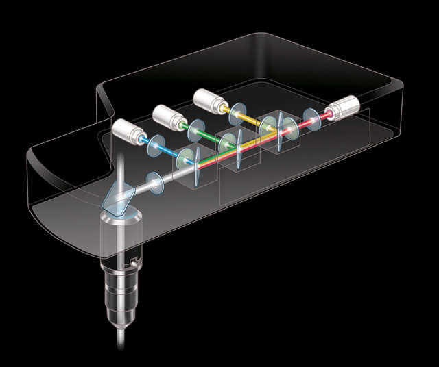
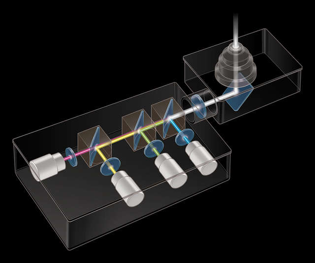
The NDDs are located as close as possible to the specimen in order to detect the maximum amount of scattered emission signals from deep within living specimens. A combination of episcopic and diascopic GaAsP NDDs for the Ni-E/FN1 upright microscope allows acquisition of bright and high S/N ratio images, by detecting both reflected and transmitted fluorescence signals.
4-channel episcopic GaAsP NDD
4-channel diascopic GaAsP NDD
Simultaneous excitation imaging with two-wavelength IR laser
The A1 MP+ and A1R MP+ are available for systems compatible with two-wavelength IR laser simultaneous excitation.
Combining the system with a femtosecond IR pulse laser with two-wavelength simultaneous output (main tunable output of 700 - 1300 nm and auxiliary fixed output of 1040 nm), enables the simultaneous excitation and imaging of two different dyes in the deep area within living cells.
Simultaneous excitation imaging with two-wavelength IR laser
Three dimensional images of 1 dpf zebrafish transgenic line, Tg[h2afv:GFP; EF1α: mCherry-zGem]. After breeding under the treatment of Phenylthiourea (PTU), which inhibits melanin synthesis, whole body was clarified with optical clearing solution LUCID-A. This transgenic line visualizes proliferating cells and chromatin with mCherry (red) and GFP (green), respectively.
Excitation wavelength: 900 nm and 1040 nm
Objective: CFI75 Apochromat 25XC W 1300 (NA 1.10, WD 2.0)
Photos courtesy of: Drs. Toshiaki Mochizuki and Ichiro Masai, Developmental Neurobiology Unit, Okinawa Institute of Science and Technology Graduate University
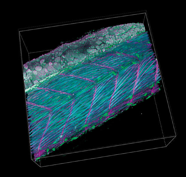
Lateral view of trunk of zebrafish transgenic line Tg[h2afv:GFP; EF1α: mCherry-CAAX] at 34 hpf. After breeding under the treatment of Phenylthiourea (PTU), which inhibits melanin synthesis, whole body was clarified with optical clearing solution LUCID-A. This transgenic line visualizes cell membrane and chromatin with mCherry (purple) and GFP (green), respectively. SHG (blue) indicates muscle fibers.
Excitation wavelength: 900 nm for SHG, GFP and 1040 nm for mCherry
Objective: CFI75 Apochromat 25XC W 1300 (NA 1.10, WD 2.0)
Photos courtesy of: Drs. Toshiaki Mochizuki and Ichiro Masai, Developmental Neurobiology Unit, Okinawa Institute of Science and Technology Graduate Universit
Ultrafast and high definition imaging with a resonant scanner [APPLIES TO A1R MP ONLY]
The A1R MP+'s resonant scanner has an ultrahigh resonance frequency of 7.8 kHz, allowing ultrafast imaging of up to 720 fps (512 x 16 pixels). Nikon's optical pixel clock generation system ensures stable, geometrically correct and evenly illuminated imaging, even at high speeds. This enables the successful visualization of in vivo rapid changes, such as reactions in living organisms, dynamics and cell interactions.
Blood cells in blood vessels within a living organism were excited by a femtosecond pulsed IR laser with the A1R MP+'s ultrahigh-speed resonant scanner, and their movements were simultaneously captured in three successive fluorescence images at 30 fps (30 msec), with three separate color channels.
Three fluorescent probes are simultaneously excited and imaged?nucleus (blue), endothelium (green), and plasma (red). The long-wavelength ultrafast laser in combination with the ultrahigh-speed resonant scanner effectively reduces photodamage and makes time resolved multiphoton imaging of biomolecules possible.
Image resolution: 512 x 512 pixels, Image acquisition speed: 30 fps, Objective: water immersion objective 60X
Photos courtesy of: Dr. Satoshi Nishimura, Center for Molecular Medicine, Jichi Medical University
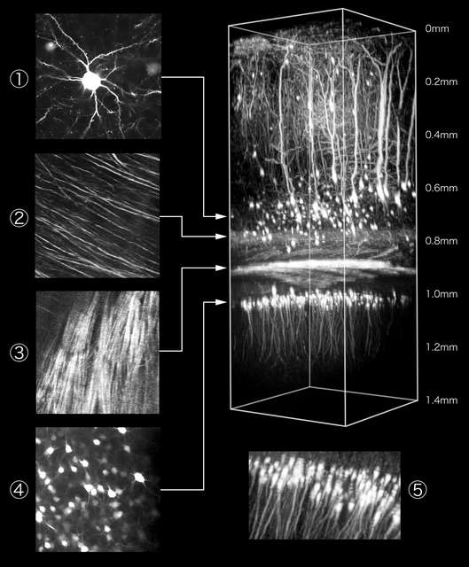
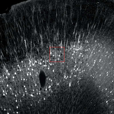
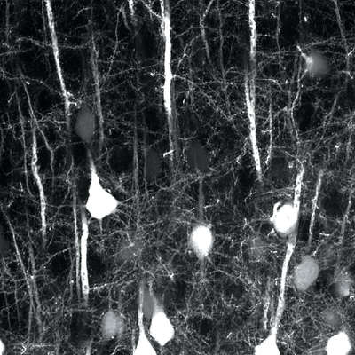
High resolution 1K image in large FOV
The resonant scanner provides finely detailed images with a maximum resolution of 1024 x 1024 pixels (15 fps). Its newly-developed sampling method produces sharper images in any configuration - even at lower resolution settings. Its large field of view (FOV18) delivers higher throughput in various imaging applications. When combined with Nikon’s high-NA objective lenses, it can achieve absolute optical precision.
Comparison of a large FOV image and detailed image of fine structures in a cleared* 2 mm brain slice of H-line mouse.
Photographed with the cooperation of: Drs. Ryosuke Kawakami, Kohei Otomo, and Tomoni Nemoto, Research Institute for Electronic Science, Hokkaido University
In vivo, large FOV, two-photon/SHG imaging of Ca2+ oscillations and collagen fibers of living pancreatic acini in an anesthetized mouse after agonist stimulation. Cyan: SHG signals, Green: GCaMP7.
Sample: GLT1-GCaMP7 mouse (G7NG817)
Microscope: A1R MP+
Objective: CFI Apochromat LWD Lambda S 20XC WI
Photo courtesy of: Ms. Yumi Yamanaka, Graduate School of Information Science and Technology, Hokkaido University, Dr. Kohei Otomo, Research Institute of Electronic Sciences, Hokkaido University, Dr. Hajime Hirase, RIKEN Brain Science Institute, Dr. Tomomi Nemoto, Research Institute of Electronic Sciences, Hokkaido University
Comparison of a large FOV image and detailed image of fine structures in a cleared* 2 mm brain slice of H-line mouse.
Photographed with the cooperation of: Drs. Ryosuke Kawakami, Kohei Otomo, and Tomoni Nemoto, Research Institute for Electronic Science, Hokkaido University
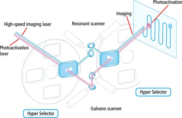
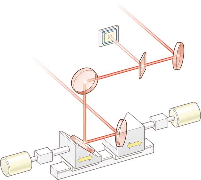
Auto laser alignment when changing multiphoton excitation wavelength
When the multiphoton laser wavelength or group velocity dispersion pre-compensation is changed, the multiphoton laser beam positional pointing at the objective back aperture may also change, resulting in uneven intensity across the image, or a slight misalignment between the IR and visible laser light paths.
Verifying the IR laser beam pointing and setting the alignment has traditionally been difficult. Nikon's A1 MP+/A1R MP+ series' auto laser alignment function, housed in the Incident Optical Unit for the multiphoton excitation light path, automatically maximizes IR laser alignments with a single click in NIS-Elements C.
(Auto laser alignment is possible within the 800 nm - 1300 nm wavelength range)