Á¤¸³Çö¹Ì°æ

Ci-E/Ci-L/Ci-S
Á¤¸³Çö¹Ì°æ > Ci-E/Ci-L/Ci-S

Ni-E Ni-U Ci-E/Ci-L/Ci-S FN1 Si Ei

Clinical and laboratory microscopes delivering unparalleled ergonomic benefits with unsurpassed optical quality.

Ci-E

Ci-L

Ci-S

ÁÖ¿ä Ư¡µé

Motorized magnification switching
Magnification can be easily changed during observation with the touch of a button on the ECLIPSE Ci-E microscope body. The buttons can also be programmed for specific objective lenses for easy switching between specific pairs. User-defined light intensity for each magnification is automatically saved and reproduced when magnification is changed. The remote control pad can also be used to easily change magnifications.
Bright and uniform Eco-illumination
The Eco-illumination* is a low-power-consuming, eco-friendly illumination system that produces uniform brightness and reduces the cost and effort of lamp replacement, thanks to its 60,000 hour, high-luminescent LED. By combining a collimator lens, fly-eye optics and LED illumination, bright and edge-to-edge uniform images can be obtained even at high magnifications. The LED illuminator features low-heat generation and provides the same color temperature at every magnification.
* Halogen model (Ci-S) is also available
Enhanced operational ease
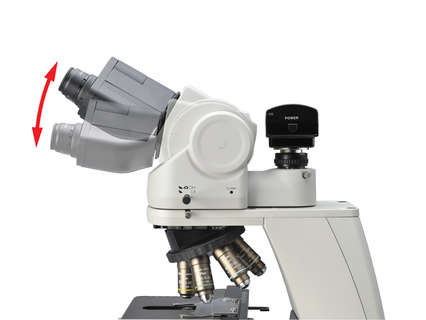
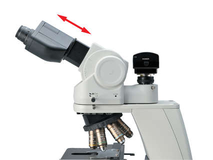
Observation with a natural posture
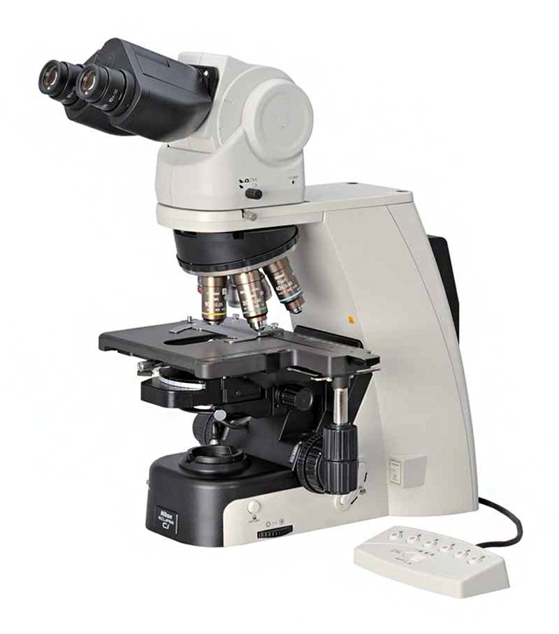
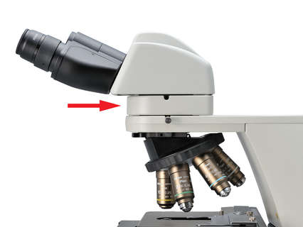
Using the ergonomic binocular tube, which features an eyepiece that can be inclined from 10¡Æ to 30¡Æand extended up to 40 mm, the microscope can be adjusted for natural posture. The eyelevel riser lifts the eyepiece tube in 25 mm increments (up to 100 mm*) and can easily be adapted to suit users with different eye-point heights.
* Up to 50 mm with ergonomic binocular tube.
User-friendly stage operation
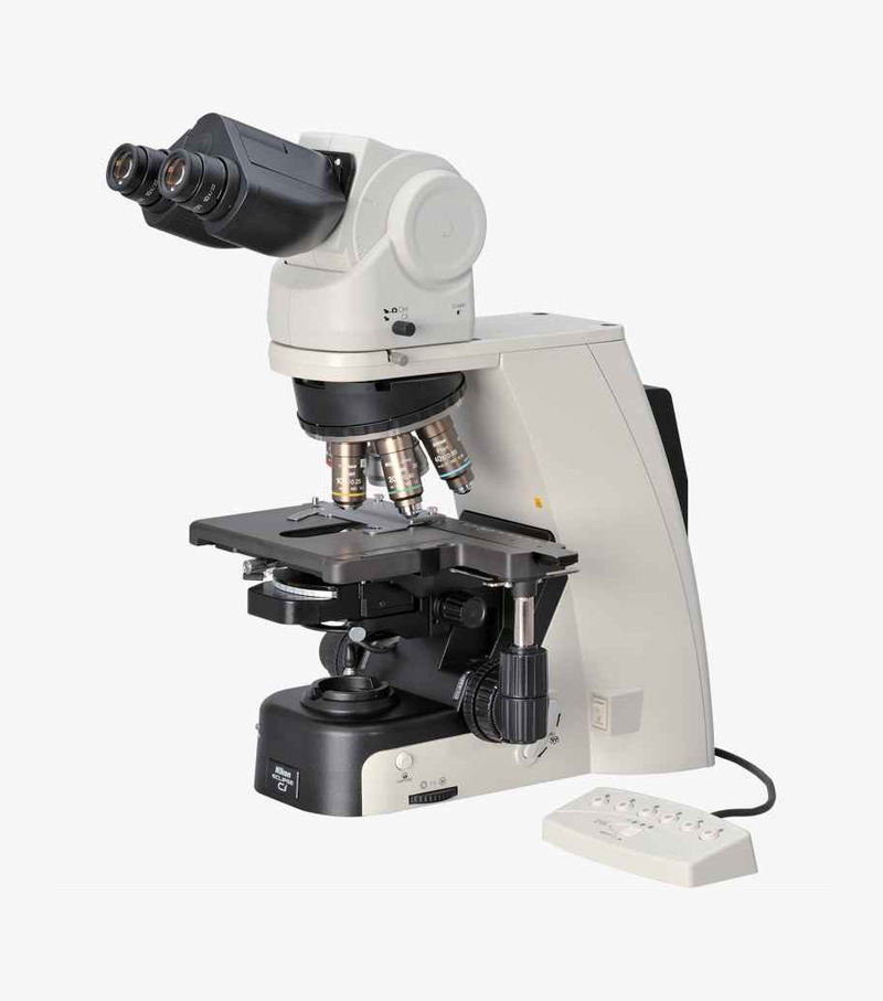
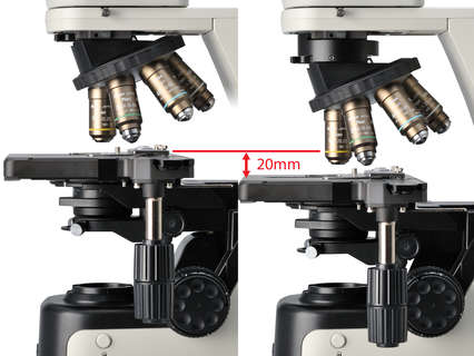
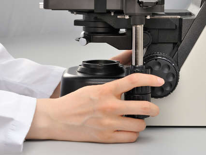
With the addition of a nosepiece spacer, the stage height can be lowered 20 mm from the standard position, reducing strain during frequent specimen change. The stage handle height can also be changed to ensure a comfortable hand position. The stage height can be locked using the refocusing knob, allowing quick refocusing after specimen changes. The stage is coated with a high-durability, scratch-resistant ceramic coating.
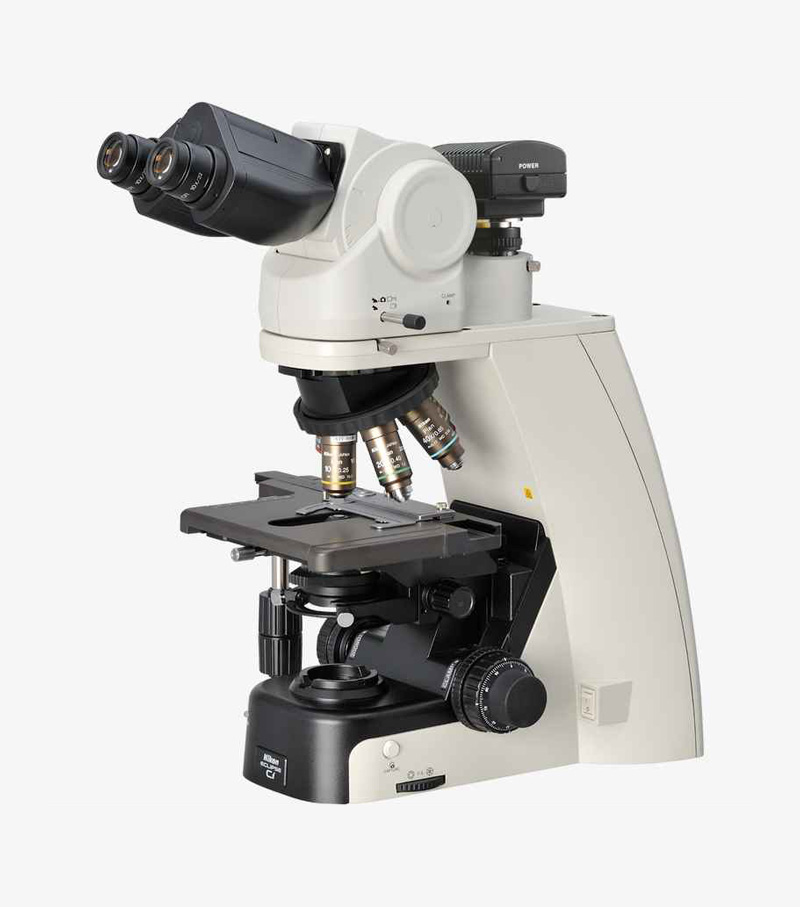
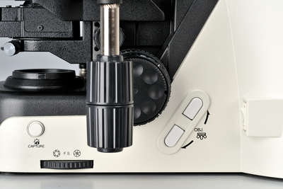
Effortless image capturing
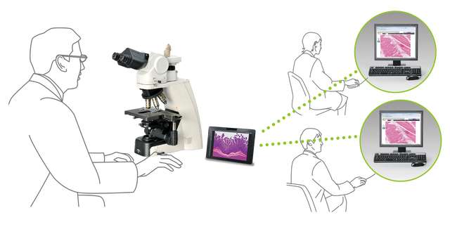
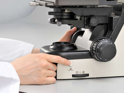
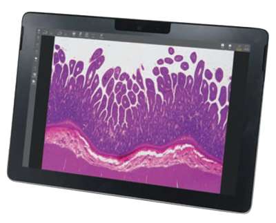
One simple click of the image capture button on the microscope base during observation enables the Digital Sight camera to capture the specimen image. Camera control software for tablet PC NIS-Elements L is equipped with a scene mode that can automatically set the optimum shooting conditions for each observation method. Since it has a network function, you can share images with a remote PC.
* NIS-Elements L is not for clinical diagnostic use.
Versatile observation techniques
Phase contrast
High-contrast images with neutral background coloration regardless of the magnification range can be captured. This observation technique is suitable for observation of unstained structures.
Simple polarizing
Ideal for observing bi-refringent samples such as collagen, amyloids and crystals.
* Two types of analyzer are available: intermediate tube type and nosepiece slider type.
Darkfield
Enables clear observation of blood or minute structures such as flagella. Dry- and oil-type condensers are available. An expander lens is utilized for brighter imaging.
Epi-fluorescence
Compact epi-fluorescence attachments that utilize noise terminating mechanisms allow weakly fluorescing specimens to be captured with great clarity and brightness. Both CI-FL epi-fluorescence attachment (incorporates up to 4 filter cubes) and D-FL epi-fluorescence attachment (incorporates up to 6 filter cubes) allow easy switching of filter cubes. High-optical-performance objective lenses for epi-fluorescence imaging, including the CFI Plan Apochromat Lambda series and the CFI Plan Fluor series, are available.