High sensitivity
Detects even faint fluorescent signals
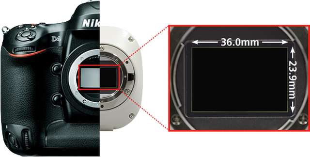
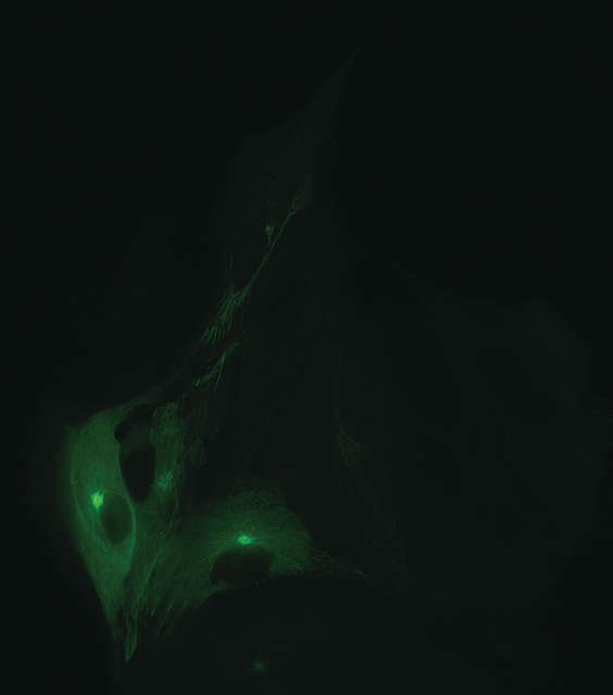
7.3μm pixels, high quantum efficiency, and very low read noise allow the DS-Qi2 to even read faint fluorescent signals.
Excellent linearity
Reliable quantitative analysis made possible
With a linearity error of ±1%, the DS-Qi2 is a superb tool for measuring intensities in fluorescence samples, including time-based intensity measurement and ratiometric measurement.
Low noise
Acquires dim fluorescent signals with ultra-low noise
2.2 electrons read noise, and this, coupled with a large full-well capacity, allows the acquisition of fluorescence images with very little noise and a very high dynamic range.
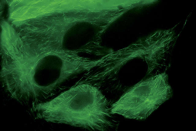
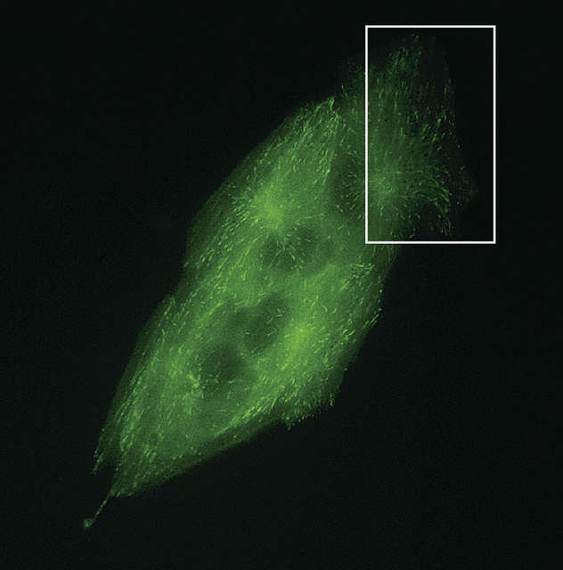
LLC-PK1 cells expressing GFP-EB3 tubulin Low noise and large linear full well capacity allows the acquisition of large dynamic range in a single capture.
A captured image is shown intensity scaled to show both the brightest and dimmest areas.
Sample courtesy of: Michael Davidson, National High Magnetic Field Laboratory, Florida State University
Time-lapse photography
Fluorescent time-lapse imaging through integration with NIS-Elements software
With a large field of view and pixel density, and low noise, the DS-Qi2 is ideal for time-resolved imaging applications.
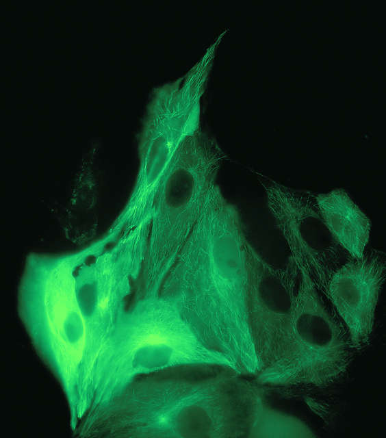
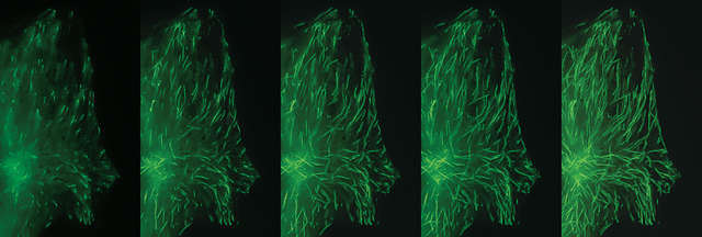
Time-lapse images (every 1 second) of LLC-PK1 cells with GFP-EB3 tubulin. Each image represents the maximum intensity projection of the timelapse, allowing visualization of the end-binding protein located on the microtubule plus-ends, and allowing tracing of the microtubule path.
DS-Qi2 captures an extremely large field of view, but still represents very fine details as demonstrated in this cropped timelapse sequence from a large FOV image.
Objective: CFI Plan Apochromat Lambda 60X Oil (NA: 1.4)
Sample courtesy of: Michael Davidson, National High Magnetic Field Laboratory, Florida State University